Bone is solid living tissue that constantly replaces itself: old bone is absorbed as new bone is made. When bone loss begins to outpace replacement, you have a condition called osteoporosis, which means porous bone. Osteoporotic bones are more vulnerable to fracture.
While breaking a bone in high school isn’t usually that big a deal, breaking a bone during middle age and beyond can be quite serious, especially when it occurs in one of the more vulnerable areas, such as the hip or spine. Osteoporotic vertebral bones can lead to a rounded back and hunched posture. And broken bones can lead to ongoing pain and dysfunction. Certain fractures, such as a broken hip, can increase one’s risk of mortality.
Women tend to develop osteoporosis earlier and more often than men (20% vs. 5%), but recent data suggests that men are more likely to die from complications related to hip fracture, making osteoporosis something that affects nearly everyone who is advancing in years.
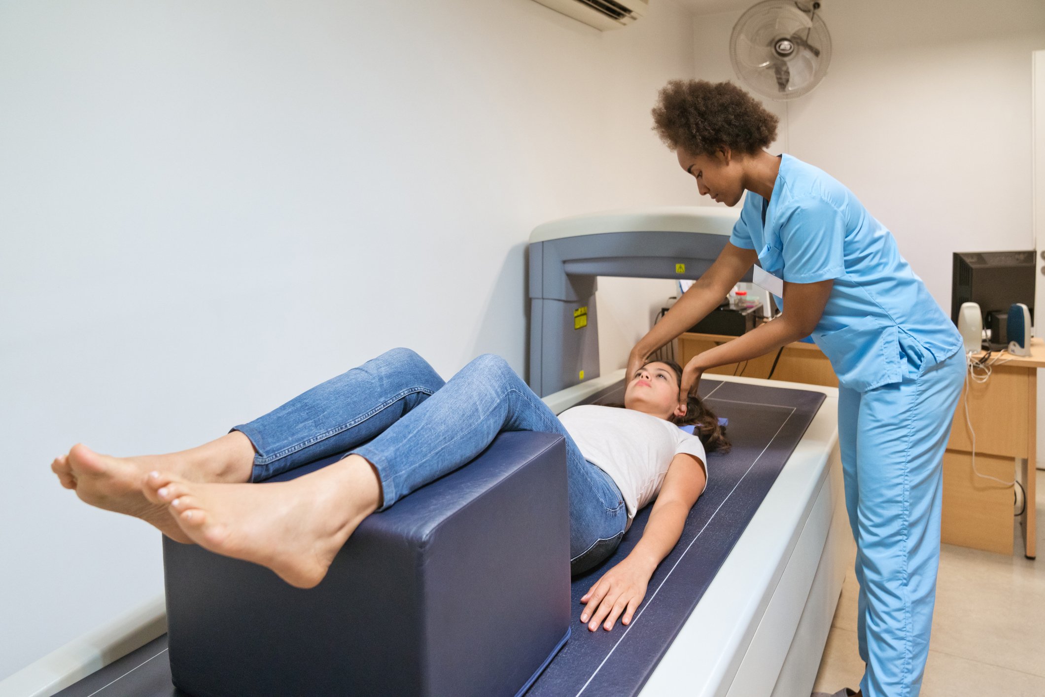
Dual-energy X-ray absorptiometry, or DEXA, is a low-dose radiation scan of the bones, usually the hip and spine, to measure their mineral content, density and strength. By examining the structure of these bones, your radiologist and referring clinician can diagnose pre-osteoporosis or osteoporosis, and assess your risk of fracture. DEXA can help discover osteoporosis early, so treatments and lifestyle changes can be employed to help prevent fracture from happening.
Who Can Benefit From a DEXA Scan?
Osteoporosis is a stealthy disease, gradually, painlessly depleting bones without making itself known until a person breaks a bone after a minor accident or notices they have gotten shorter.
For the general population at average risk, DEXA is recommended for:
Women 65 and older
Men 70 and older
A supplementary exam is often ordered within the following two years to monitor changes and gauge the effectiveness of any treatment. In rare cases, patients taking certain medications, like high-dose steroids, may need a follow-up scan as soon as six months.
People at increased risk may benefit from DEXA as early as age 50. Elevated risk factors include:
Age 50 and older and suffered a broken bone
Age 50 and older and lost more than an inch in height
Additional risk factors to consider when talking to your clinician include:
Personal or parental history of fracture
Caucasian or Asian race
Cigarette smoking
Heavy drinking
Early menopause (before age 45)
Post-menopausal and not taking estrogen
Sedentary lifestyle
Being underweight
Poor diet
High bone turnover
Type 1 diabetes
Osteoarthritis
Rheumatoid arthritis
Thyroid or parathyroid condition
Use of corticosteroids, anti-seizure drugs or barbiturates
How Is DEXA Performed?
DEXA is quick, easy, noninvasive and completely painless.
First, your technologist will have you lie back on a specialized x-ray table and help position you for optimal imaging.
You lie still while the scanner passes over the area being imaged. This typically takes about 10-20 minutes total.
Images are sent from the scanner to a computer monitor.
Your DEXA-trained radiologist examines and interprets the images, and shares them with your referring clinician.
Your clinician discusses the results with you, and, if necessary, recommends a treatment protocol.
How Is Bone Density Measured?
DEXA produces two scores, T and Z, which are taken together to assess bone density and strength.
The T score compares your bone mass to that of a young healthy adult of the same gender.
T score: -1.0 and above = normal bone mass
T score: -1.1 – -2.4 = low bone mass (osteopenia, or pre-osteoporosis)
T score: -2.5 or less = osteoporosis
The Z score compares the amount of bone you have to average people of the same gender and general age and height. A Z score below -2.0 indicates below average bone mass, and may show that you are losing bone more rapidly than is normal for someone in your age group.
Your radiologist and referring clinician will use your score to estimate your risk of suffering a fracture and determine if preventive treatment is needed.
Is DEXA Safe?
The dose of ionizing radiation produced by a DEXA scan is very small, about 1/10th that of a CT scan. DEXA is considered safe and has been FDA-approved for clinical use since 1988.
Treatment for Osteoporosis
Your clinician will review the results of your DEXA scan and your radiologist’s report, and suggest next steps based on your specific situation. Treatments for osteoporosis include bone-building medications, weight-bearing exercises, diet changes and certain vitamin and mineral supplements.
The earlier osteoporosis is discovered, the sooner effective therapies can be initiated to help prevent or slow further bone loss, reducing the risk of fracture.
If you have one or more risk factors for osteoporosis, talk to your clinician about whether a DEXA scan might be right for you.
RAO offers DEXA testing at our Women’s Imaging Center and TimberRidge Imaging Center. Beginning in March, 2023, we will offer DEXA at our new TimberRidge Imaging Center at Heathbrook Pavillion.


